back to texbook back to laboratory exercsise
Neutron activation analysis (NAA) is a sensitive analytical technique for performing both qualitative and quantitative multi-element analysis of major, minor and trace elements in samples of almost every conceivable field of scientific or technical interest. For many elements and applications, NAA offers sensitivities that are superior to those attainable by other methods. In addition, because of its accuracy and reliability, NAA is generally recognized, as the "referee method" of choice when new procedures are being developed or when other methods yield results that do not agree. NAA is widespread worldwide at most nuclear reactor installations.
Neutron activation analysis was discovered in 1936 when Hevesy and Levi found that samples containing certain rare earth elements became highly radioactive after exposure to a source of neutrons. From this observation, they quickly recognized the potential of employing nuclear reactions on samples followed by measurement of the induced radioactivity to facilitate both qualitative and quantitative identification of the elements present in the samples.
The basic essentials required to carry out an analysis of samples by NAA are a source of neutrons, instrumentation suitable for detecting gamma rays, and a detailed knowledge of the reactions that occur when neutrons interact with target nuclei. Brief descriptions of the NAA method, reactor neutron sources, and gamma-ray detection are given below.
The NAA Method
The sequence of events occurring during the most common type of nuclear reaction used for NAA, namely the neutron capture or (n,γ) reaction, is illustrated in the figure below.| FIG 1:Diagram illustrating the process of neutron capture by a target nucleus followed by the emission of gamma rays. |
When a neutron interacts with the target nucleus via a non-elastic collision, a compound nucleus forms in an excited state. The excitation energy of the compound nucleus is due to the binding energy of the neutron within the nucleus. The compound nucleus will almost instantaneously de-excite into a more stable configuration through emission of one or more characteristic prompt gamma rays. In many cases, this new configuration yields
radioactive nucleus which also de-excites (or decays) by emission of one or more characteristic delayed gamma rays, but at a much slower rate according to the unique half-life of the radio-active nucleus. Depending upon the particular radioactive species, half-lives can range from fractions of a second to several years.
In principle, therefore, with respect to the time of measurement, NAA falls into two categories: (1) prompt gamma-ray neutron activation analysis (PGNAA), where measurements take place during irradiation, or (2) delayed gamma-ray neutron activation analysis (DGNAA), where the measurements follow radioactive decay. The latter operational mode is more common; thus, when one mentions NAA it is generally assumed that measurement of the delayed gamma rays is intended. About 70% of the elements have properties suitable for measurement by NAA.
Neutrons
Although there are several types of neutron sources (reactors, accelerators, and radioisotopic neutron emitters) one can use for NAA, nuclear reactors with their high fluxes of neutrons from uranium fission offer the highest available sensitivities for most elements. Different types of reactors and different positions within a reactor can vary considerably with regard to their neutron energy distributions and fluxes due to the materials used to moderate (or reduce the energies of) the primary fission neutrons. However, as shown in the figure below, most neutron energy distributions are quite broad and consist of three principal components (thermal, epithermal, and fast).
 |
| Fig 2:A typical reactor neutron energy spectrum showing the various components used to describe the neutron energy regions. |
The thermal neutron component consists of low-energy neutrons (energies below 0.5 eV) in thermal equilibrium with atoms in the reactor's moderator. At room temperature, the energy spectrum of thermal neutrons is best described by a Maxwell-Boltzmann distribution with a mean energy of 0.025 eV and a most probable velocity of 2200 m/s. In most reactor irradiation positions, 90-95% of the neutrons that bombard a sample are thermal neutrons. In general, a one-megawatt reactor has a peak thermal neutron flux of approximately 1 neutrons per square centimeter per second.
The epithermal neutron component consists of neutrons (energies from 0.5 eV to about 0.5 MeV) which have been only partially moderated. A cadmium foil 1 mm thick absorbs all thermal neutrons but will allow epithermal and fast neutrons above 0.5 eV in energy to pass through. In a typical unshielded reactor irradiation position, the epithermal neutron flux represents about 2% the total neutron flux. Both thermal and epithermal neutrons induce (n,γ) reactions on target nuclei. An NAA technique that employs only epithermal neutrons to induce (n,γ) reactions by irradiating the samples being analyzed inside either cadmium or boron shields is called epithermal neutron activation analysis (ENAA).
The fast neutron component of the neutron spectrum (energies above 0.5 MeV) consists of the primary fission neutrons, which still have much of their original energy following fission. Fast neutrons contribute very little to the (n,γ) reaction, but instead induce nuclear reactions where the ejection of one or more nuclear particles - (n,p), (n,n'), and (n,2n) - are prevalent. In a typical reactor irradiation position, about 5% of the total flux consists of fast neutrons. An NAA technique that employs nuclear reactions induced by fast neutrons is called fast neutron activation analysis (FNAA).
The neutron source in this exercise is the JEEP II reactor at Institute for Energy Technology (IFE) at Kjeller. It is a heavy water moderated reactor with a thermal power of 2 MW, and the thermal neutron flux in the irradiation position is about 1.5•1013n•cm-2•s-1.
The NAA-laboratory at IFE is equipped with a “rabbit” system, which transports the sample pneumatically from the laboratory into the irradiation position. After a pre-programmed irradiation time the sample is returned pneumatically to the NAA-laboratory. The transfer time is 25-30 s over a distance of 200 m. This rabbit system is used for short irradiation times (< 3 h). For longer irradiation times the samples are loaded manually by a loading machine vertically from the reactor top.
Promt vs Delayed NAA
As mentioned earlier, the NAA technique can be categorized according to whether gamma rays are measured during neutron irradiation (PGNAA = Prompt-Gamma Neutron Activation Analysis) or at some time after the end of the irradiation (DGNAA = Delayed-Gamma Neutron Activation Analysis). The PGNAA technique is generally performed by using a beam of neutrons extracted through a reactor beam port. Fluxes on samples irradiated in beams are on the order of one million times lower than on samples inside a reactor but detectors can be placed very close to the sample compensating for much of the loss in sensitivity due to flux. The PGNAA technique is most applicable to elements with extremely high neutron capture cross-sections (B, Cd, Sm, and Gd); elements which decay too rapidly to be measured by DGNAA; elements that produce only stable isotopes; or elements with weak decay gamma-ray intensities.
DGNAA (sometimes called conventional NAA) is useful for the vast majority of elements that produce radioactive nuclides. The technique is flexible with respect to time such that the sensitivity for a long-lived radionuclide that suffers from interference by a shorter-lived radionuclide can be improved by waiting for the short-lived radionuclide to decay. This selectivity is a key advantage of DGNAA over other analytical methods.
Instrumental vs Radiochemical NAA
With the use of automated sample handling, gamma-ray measurement with solid-state detectors, and computerized data processing it is generally possible to simultaneously measure more than thirty elements in most sample types without chemical processing. The application of purely instrumental procedures is commonly called instrumental neutron activation analysis (INAA) and is one of NAA's most important advantages over other analytical techniques. If chemical separations are done to samples after irradiation to remove interferences or to concentrate the radioisotope of interest, the technique is called radiochemical neutron activation analysis (RNAA). The latter technique is performed infrequently due to its high labor cost.
Measurement and Gamma-rays
The instrumentation used to measure gamma rays from radioactive samples generally consists of a semiconductor detector, associated electronics, and a computer-based, multi-channel analyzer (MCA/computer). Most NAA labs operate one or more hyperpure or intrinsic germanium (HPGe) detectors, which operate at liquid nitrogen temperatures (77 degrees K) by mounting the germanium crystal in a vacuum cryostat, thermally connected to a copper rod or "cold finger". Although HPGe detectors come in many different designs and sizes, the most common type of detector is the coaxial detector, which in NAA is useful for measurement of gamma-rays with energies over the range from about 50 keV to 3.0 MeV.
The two most important performance characteristics requiring consideration when purchasing a new HPGe detector are energy resolution and detection efficiency. Other characteristics to consider are peak shape, peak-to-Compton ratio, crystal dimensions or shape, and price.
The detector's energy resolution is a measure of its ability to separate closely spaced peaks in a gamma spectrum. In general, detector resolution is specified in terms of the full width at half maximum (FWHM) of the 122-keV photopeak of 57Co and the 1332-keV photopeak of 60Co. For most NAA applications, a detector with 1.0-keV resolution or below at 122 keV and 1.8 keV or below at 1332 keV is sufficient.
 |
 |
|---|---|
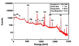
| Fig 3: Gamma-ray spectrum showing several short-lived elements measured in a sample of pottery irradiated for 5 seconds, decayed for 25 minutes, and counted for 12 minutes with an HPGe detector. |
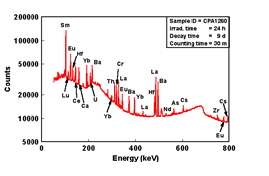
Fig 4:Gamma-ray spectrum from 0 to 800 keV showing medium- and long-lived elements measured in a sample of pottery irradiated for 24 hours, decayed for 9 days, and counted for 30 minutes on an HPGe detector. |
The true absolute “intrinsic” counting efficiency has to be measured by using certified standard gamma sources with known energies and intensities where the disintegration rate is known within narrow error limits.
Typical gamma-ray spectra from an irradiated pottery specimen are shown in figures above using two different irradiation and measurement procedures.
Using Gamma-ray Counts to Calculate Element Concentration
In principle, it is possible to perform quantitative NAA by using the general formula for activation:
Where:
w = mass of unknown element in sample
Rx = measured count rate of unknown element (cps)
M = atomic weight of unknown element
λX
decay constant of the measured radionuclide of the unknown element
ln2/T1/2Where T1/2 is the half-life of this radionuclide (s-1)td
decay time
time between irradiation end and midpoint of counting period (s)ɛx = counting efficiency of the detected gamma energy
σ = thermal neutron reaction cross section of the unknown element (barn = 10-24 cm2)
φ = neutron flux in irradiation position (ns-1cm-2).
NA = Avogadro’s number (6.023•1023)
IA = natural occurrence of the target isotope in the actual element (as fraction in the range 0-1)
ti = irradiation time (s)
However, in practice, the parameters φ and σ are not easily determined exactly. Therefore, a simpler method is to carry out so-called comparative analysis where the activity in the sample is compared to the activity in a simultaneously irradiated standard of the same element. If the unknown sample and the comparator standard are both measured on the same detector, then one needs to correct the difference in decay between the two. One usually decay corrects the measured counts (or activity) for both samples back to the end of irradiation using the half-life of the measured isotope. The following relation may be put up:

If the unknown sample and the comparator standard are both measured on the same detector, ɛxp= ɛxpand the following simple expression results:

Sensitivities Available by NAA
The sensitivities for NAA are dependent upon the irradiation parameters (i.e., neutron flux, irradiation and decay times), measurement conditions (i.e., measurement time, detector
efficiency), nuclear parameters of the elements being measured (i.e., isotope abundance, neutron cross-section, half-life, and gamma-ray abundance). The accuracy of an individual
NAA determination usually ranges between 1 to 10 percent of the reported value. Table I lists the approximate sensitivities for determination of elements assuming interference free
spectra.
Table 1. Estimated detection limits for INAA using decay gamma rays. Assuming irradiation in a reactor neutron flux of 11013 n cm-2 s-1.
| Sensitivity 10-12 g |
Element |
| <1 |
Dy, Eu |
| 1-10 |
In,Lu, Mn |
| 10-100 |
Au, Ho, Ir, Re, Sm, W |
| 100-1000 |
Ag, Ar, As, Br, Cl, Co, Cs, Cu, Er, Ga, Hf, I, La, Sb, Sc, Se, Ta, Tb, Th, Tm, U, V, Yb |
| 103-104 |
Al, Ba, Cd, Ce, Cr, Hg, Kr, Gd, Ge, Mo, Na, Nd, Ni, Os, Pd, Rb, Rh, Ru, Sr, Te, Zn, Zr |
| 104-105 |
Bi, Ca, K, Mg, P, Pt, Si, Sn, Ti, Tl, Xe, Y |
| 105-106 |
F, Fe, Nb, Ne |
| >106 |
Pb, S |
Possible Applications For NAA
A few examples of the application of NAA in various areas of technology and research are mentioned in table below:
Examples of NAA-applications in technology and research.
| Area |
Description |
| Agriculture |
Beet pulp, lipids, hay, oil, meat, soil, fertilizers etc. |
| Anthropology |
Obsidian, teeth, bones |
| Archaeology |
Pottery shards, clay samples, painting fractions, coins, jewelery etc. |
| Biology |
Chemicals, sugar, enzymes, solutions, ants |
| Botany |
Wheat spores, plant physiology |
| Chemistry |
Contaminants in oxides, salts, pure crystals, metal residue, "pure" liquids etc. |
| Engineering |
Corrosion and wear |
| Environment |
Precipitations, leak-offs, air samples (incl. particles), water samples, plants |
| Fisheries |
Fish and seafood, fish shells and bones |
| Forensics |
Bullets, paint, glass, metals, gunshot residues, hair and nails etc. |
| Forestry |
Wood rings, Tree needles, soil |
| Geology |
Minerals, meteorites, gemstones |
| Industry |
Pure metals, chemical compounds, oils, thin-film deposits alloys, rock etc. |
| Medicine |
Water, skin, skin, hair, nails, various organs, various body fluids |
| nuclear |
Fuel pin cladding, water, uranium mineral ores |
| Oceanography |
Fossils, sediments, basalts |
| Pharmacy |
Chemicals |
